Circularly Polarized Luminescence for Beginners
Circularly Polarized Luminescence for Beginners
Blog Article
Getting My Circularly Polarized Luminescence To Work
Table of ContentsThe smart Trick of Circular Dichroism That Nobody is Talking AboutThe Best Guide To Circularly Polarized LuminescenceMore About Uv/visThe Of Uv/visHow Uv/vis/nir can Save You Time, Stress, and Money.Some Known Facts About Uv/vis/nir.Excitement About Uv/vis/nirThe Ultimate Guide To Uv/vis/nirNot known Details About Uv/vis/nir What Does Circular Dichroism Mean?5 Easy Facts About Circularly Polarized Luminescence ShownUv/vis/nir Things To Know Before You Get ThisThe Of Uv/vis
It is then scanned through the sample and the referral solutions. Portions of the incident wavelengths are transferred through, or shown from, the sample and the reference. Electronic circuits convert the relative currents into linear transmission portions and/or absorbance/concentration worths.The transmission of a reference substance is set as a baseline (datum) worth, so the transmission of all other substances are recorded relative to the initial "zeroed" substance. The spectrophotometer then transforms the transmission ratio into 'absorbency', the concentration of particular components of the test sample relative to the initial substance.
Because samples in these applications are not readily available in large amounts, they are specifically fit to being evaluated in this non-destructive technique. In addition, precious sample can be conserved by using a micro-volume platform where as little as 1u, L of sample is required for total analyses. A quick explanation of the treatment of spectrophotometry consists of comparing the absorbency of a blank sample that does not contain a colored substance to a sample that contains a colored compound.
All about Circularly Polarized Luminescence
In biochemical experiments, a chemical and/or physical home is picked and the procedure that is used specifies to that property in order to derive more information about the sample, such as the quantity, pureness, enzyme activity, etc. Spectrophotometry can be used for a variety of strategies such as determining optimal wavelength absorbance of samples, figuring out ideal p, H for absorbance of samples, determining concentrations of unknown samples, and identifying the p, Ka of numerous samples.: 21119 Spectrophotometry is likewise a useful procedure for protein filtration and can also be used as an approach to produce optical assays of a compound.
It is possible to understand the concentrations of a 2 part mix using the absorption spectra of the standard services of each component. To do this, it is required to know the extinction coefficient of this mix at two wave lengths and the extinction coefficients of services that contain the recognized weights of the 2 components.
Circularly Polarized Luminescence Things To Know Before You Buy
Region. The concentration of a protein can be approximated by measuring the OD at 280 nm due to the existence of tryptophan, tyrosine and phenylalanine.
This method needs a spectrophotometer capable of determining in the UV area with quartz cuvettes.: 135 Ultraviolet-visible (UV-vis) spectroscopy includes energy levels that delight electronic shifts. Absorption of UV-vis light delights molecules that are in ground-states to their excited-states.
20. 8 O.D. Ink makers, printing business, textiles vendors, and much more, require the data supplied through colorimetry. They take readings in the area of every 520 nanometers along the noticeable area, and produce a spectral reflectance curve or an information stream for alternative discussions. These curves can be used to test a new batch of colorant to inspect if it makes a match to requirements, e.
The Single Strategy To Use For Circular Dichroism
Conventional noticeable region spectrophotometers can not detect if a colorant or the base material has fluorescence. This can make it difficult to manage color concerns if for example one or more of the printing inks is fluorescent. Where a colorant includes fluorescence, a bi-spectral fluorescent spectrophotometer is used (https://www.magcloud.com/user/olisclarity1). There are 2 significant setups for visual spectrum spectrophotometers, d/8 (spherical) and 0/45.
Scientists utilize this instrument to determine the amount of compounds in a sample. In the case of printing measurements two alternative settings are frequently used- without/with uv filter to control better the impact of uv brighteners within the paper stock.
The Best Strategy To Use For Circular Dichroism
Some applications require little volume measurements which can be performed with micro-volume platforms. As explained in the applications section, spectrophotometry can be used in both qualitative and quantitative analysis of DNA, RNA, and proteins. Qualitative analysis can be utilized and spectrophotometers are used to tape spectra of substances by scanning broad wavelength regions to identify the absorbance residential or commercial properties (the strength of the color) of the compound at each wavelength.

3 Easy Facts About Circularly Polarized Luminescence Shown
One significant factor is the type of photosensors that are available for various spectral regions, but infrared measurement is also difficult since practically everything discharges IR as thermal radiation, particularly at wavelengths beyond about 5 m. Another complication is that several products such as glass and plastic soak up infrared, making it incompatible as an optical medium.
Samples for IR spectrophotometry may be smeared in between two discs of potassium bromide or ground with potassium bromide and pressed into a pellet. Where aqueous services are to be measured, insoluble silver chloride is used to construct the cell. Spectroradiometers, which run almost like the visible region spectrophotometers, are designed to measure the spectral density of illuminants. 2013. p. 13. Allen, DW; Cooksey, C; Tsai, BK (Nov 13, 2009). "Spectrophotometry". Recovered Dec 23, 2018. Ninfa AJ, Ballou DP, Benore M (2010 ). Essential Laboratory Methods for Biochemistry and Biotechnology (second ed.). Hoboken: Wiley & Sons. ISBN 9780470087664. OCLC 488246403. Schwedt G (1997 ). The important guide to analytical chemistry.
Oke, J. B.; Gunn, J. E.
Some Ideas on Circularly Polarized Luminescence You Should Know
Ninfa AJ, Ballou DP, Benore M (2015 ). Basic Laboratory Approaches for Biochemistry and Biotechnology (3, rev. ed.). UV/Vis. Lab Devices.
Uv/vis Things To Know Before You Buy
"Applied Spectrophotometry: Analysis of a Biochemical Mixture". Biochemistry and Molecular Biology Education. Journal of Biochemistry Education.
The Ultimate Guide To Circular Dichroism
U.S. Department of Commerce National Bureau of Standards special publication; 378. Washington, D.C.: U.S. National Bureau of Standards.
The procedure starts with a controlled light that illuminates the analyzed sample. In the case of reflection, as this light communicates with the sample, some is absorbed or released. The discharged light journeys to the detector, which is evaluated, measured, and presented as industry-standard color scales and indices.
All terms are evaluated over the visible spectrum from 400 to 700 nm. In the case of transmission, when the light interacts with the sample, it is either absorbed, shown, or transferred.
The Ultimate Guide To Uv/vis/nir
Examples include APHA (American Public Health Association) for watercolor and pureness analysis, ASTM D1500 for petrochemical color analysis, edible oil indices used in food, and color analyses of drinks. All terms are evaluated over the visible spectrum from 400 to 700 nm.
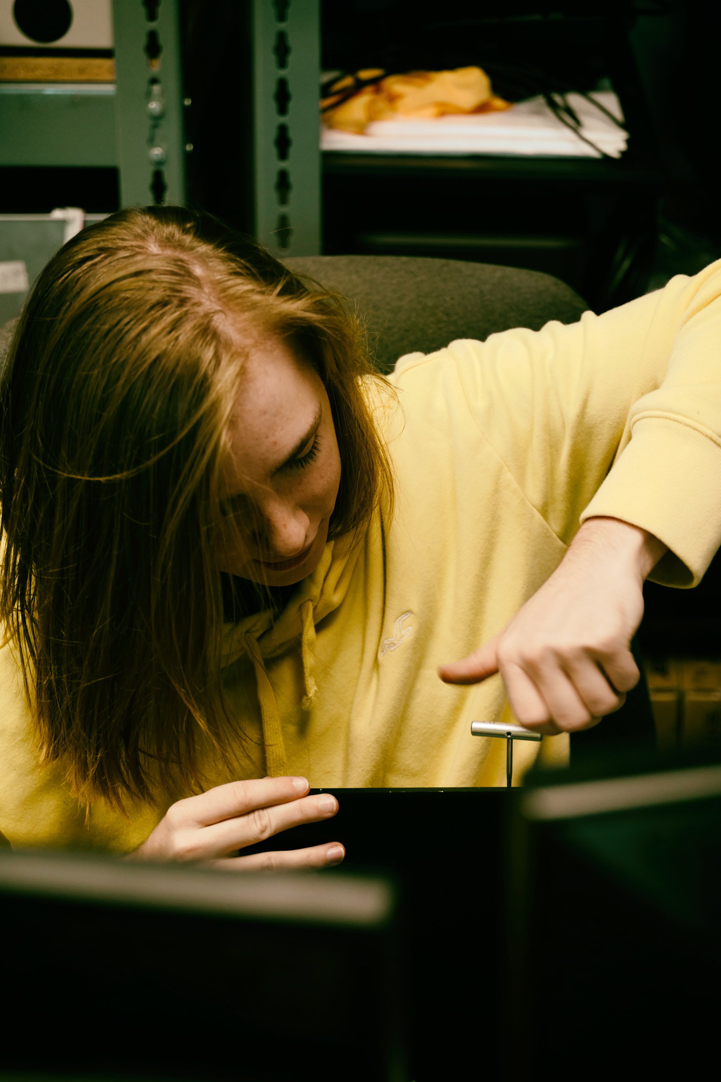
Image Credit: Matej Kastelic/ Dr. Arnold J. Beckman and his associates at the National Technologies Laboratories first created the spectrophotometer in 1940. In 1935 Beckman founded the business, and the discovery of the spectrophotometer was their most ground-breaking creation.
Some Known Facts About Circular Dichroism.
99% precision. Gradually, researchers kept improving the spectrophotometer design to improve its efficiency. The UV capabilities of the design B spectrophotometer were improved by changing the glass prism with a quartz prism. Ultimately, the Model DU was created, consisting of a hydrogen light and other enhancements. This instrument was used in commercial laboratories, clinics, and chemistry and biochemistry departments.
After 1984, double-beam versions of the device were created. The addition of external software application with the arrangement of onscreen screens of the spectra came in the 1990s. Generally, a spectrophotometer is comprised of two instruments, particularly, a spectrometer and a photometer. A fundamental spectrophotometer contains a light, a monochromator, a collimator for straight light beam transmission, a cuvette to position a sample, and a photoelectric detector.
The Only Guide for Circular Dichroism
There are various types of spectrophotometers in different shapes and sizes, each with its own function or functionality. A spectrophotometer determines how much light is shown by chemical components. circular dichroism. It determines the difference in light intensity based upon the overall amount of light presented to a sample and the amount of beam that passes through the sample option
According to the instrument's design, the sample is positioned in between the spectrometer and the photometer. After the light is gone through the sample, the photometer determines its strength and find out here now displays the reading. A spectrophotometer is used to identify the concentration of both colorless and colored solutes in an option. This instrument is used to determine the rate of a reaction.
Report this page